Source: Advanced Science, written by Sarah Alexandrou; Kristine J Fernandez; Min Li Huang; Ewan K A Millar; Claire Vennin; Leila Eshraghi; Tan Dat Timothy Lam; Victoria Lee; Nyah C Brooks; Janett Stoehr; Cristian Guaman Mendez; Douglas Strathdee; David Stevenson; David Herrmann; Sandra O'Toole; Paul Timpson; Max Nobis; C Elizabeth Caldon (October 11, 2026). AI-generated summary by biochip.com, published . Not independently reviewed.
Key takeaways
- A Cdk1-FRET biosensor mouse visualized rising Cdk1 activity as mammary tumors progressed to invasive ductal carcinoma.
- NeoPalAna nonresponders incompletely suppressed CDK1, while resistant cells, mouse tumors, and patient tumors showed elevated CDK1 activity.
- CDK1 is a candidate biomarker and requires prospective clinical validation before it can guide patient treatment decisions.
Researchers at UNSW Sydney, the Garvan Institute of Medical Research, and collaborating centers created a Cdk1-FRET biosensor mouse that lets them watch a key cell-division enzyme switch on inside developing breast tumors. They paired this real-time imaging platform with patient cohorts and treatment models to examine whether CDK1 activity signals aggressive estrogen receptor-positive breast cancer and resistance to CDK4/6 inhibitors. The team found that CDK1 activity rose as tumors progressed to invasive ductal carcinoma and rebounded rapidly after initial drug suppression in experimental models. The work identifies CDK1 as a candidate biomarker, although it does not establish a clinical test ready for patient care.
A fluorescent window into cell division
CDK1, short for cyclin-dependent kinase 1, is an enzyme that helps cells enter mitosis, the stage when one cell divides into two. Think of it as a final green light in the cell-division process: upstream signals may slow traffic, but active CDK1 can help a cell proceed toward division.
The new mouse model uses Förster resonance energy transfer, or FRET, a fluorescence method that changes its optical signal when molecules move close together. The Cdk1-FRET sensor converts CDK1 activity into a measurable light signal, allowing researchers to visualize enzyme dynamics within living tissue rather than relying only on fixed tumor samples.
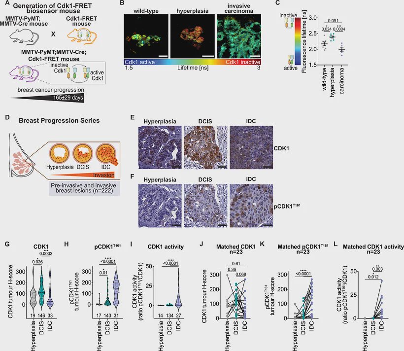
The team crossed the biosensor mouse with the MMTV-PyMT breast cancer model and an MMTV-Cre line that activates the sensor in mammary tissue. Using this system, they visualized increasing Cdk1 activity as tumors transitioned to invasive ductal carcinoma, a form of breast cancer that has spread beyond the milk ducts into surrounding tissue.
Signals of invasive disease and relapse risk
The clinical analyses focused on estrogen receptor-positive, or ER-positive, breast cancer. This is the most common breast cancer subtype and is typically treated with endocrine therapy, which blocks hormone signaling that can fuel tumor growth.
In patient cohorts, the researchers reported that pCDK1 T161, a chemically modified form of CDK1 associated with activity, was specifically increased in invasive ductal carcinoma. They also found that pCDK1 Y15 was associated with poor relapse-free survival in stage 1 disease.
Phosphorylation, the attachment of a small phosphate group to a protein, can alter how an enzyme behaves. Measuring these CDK1 phosphorylation sites may therefore provide more functional information than measuring total CDK1 gene or protein levels alone.
Watching tumors adapt to CDK4/6 inhibitors
CDK4/6 inhibitors, including palbociclib, ribociclib, and abemaciclib, are designed to stop cancer cells before they copy their DNA and divide. Yet clinicians still lack standard molecular markers that reliably identify which high-risk ER-positive tumors will respond durably.
The researchers found that the CDK1 and cyclin B1 complex was initially suppressed by CDK4/6 inhibition but rebounded within three days. In samples from the NeoPalAna trial, tumors that did not respond to treatment showed incomplete CDK1 suppression.
The same pattern appeared across experimental resistance models. Palbociclib-resistant MCF-7 breast cancer cells regained baseline CDK1 activity, nonresponsive tumors had elevated Cdk1 activity in mice, and patient tumors resistant to CDK4/6 inhibitors also showed elevated CDK1.
Why This Matters
Current treatment decisions for high-risk early ER-positive breast cancer use clinical features such as tumor size, grade, and lymph-node involvement. Ki67, another marker of cell proliferation, has had inconsistent laboratory reliability and was removed as a required selection criterion for adjuvant abemaciclib treatment.
The Cdk1-FRET mouse offers a way to study the timing of treatment adaptation, not simply whether a tumor contains a marker at one moment. That distinction matters because the reported rebound in CDK1 activity suggests tumors can re-establish a route to cell division soon after CDK4/6 inhibition begins.
Next, CDK1-based measurements will need prospective clinical testing to determine whether they can guide treatment choices, predict recurrence, or identify resistance early enough to change care.
Topics: Biosensors · Cancer
Start here: Biosensors explained
Related articles
- Liquid Biopsy in Breast Cancer: Biomarkers and Clinical Applications
- Development of semiconductor microchip that can detect prostate cancer markers with ultra-high sensitivity
- Engineered Allosteric Biosensor for In Situ DNA Glycosylase Detection in Neuroblastoma Risk Stratification and Drug Resistance Monitoring
- Screening Colorectal Tumor-Associated Fibroblasts With a Dual-Channel THz Biosensor