Source: Drug Today, by Drug Today Bureau (September 30, 2026). AI-generated summary by biochip.com, published . Not independently reviewed.
Key takeaways
- Taiyuan University of Technology and Shanxi Bethune Hospital developed a spiral chip that recovered 89.2% of MCF-7 tumor cells.
- A YOLOv8 image model identified tumor cells with 96.0% accuracy using bright-field microscope images.
- The system used artificial samples with cell lines, not patient blood, and is not yet a clinically validated liquid-biopsy test.
Researchers at Taiyuan University of Technology and Shanxi Bethune Hospital have paired a spiral cell-sorting chip with artificial intelligence to find rare tumor cells in blood samples. The system uses inertial microfluidics, the controlled movement of cells through tiny fluid channels, to concentrate larger tumor cells before software examines microscope images. In laboratory-made samples, it recovered 89.2% of MCF-7 breast cancer cells and identified tumor cells with 96.0% accuracy. The work could simplify an important step in liquid biopsy research, though it has not yet been validated using blood from cancer patients.
Finding a Few Cells in a Biological Haystack
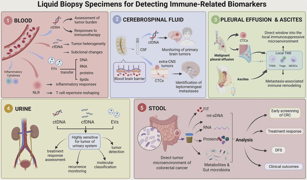
Circulating tumor cells, or CTCs, are cells that detach from a tumor and enter the bloodstream. They may offer clues about a cancer's progression, but they are exceptionally difficult to find: a sample can contain only one to ten CTCs among roughly one billion blood cells.
That makes CTC analysis a two-part problem. A system must first separate the rare target cells from the overwhelming number of blood cells, then determine whether the remaining cells are actually cancerous.
A Spiral Chip Sorts Cells by Physical Traits
Prof. Xiaochun Li's team built a spiral microfluidic chip that sorts cells according to physical properties, including size. Think of it as a very small, curved water slide: as fluid moves through the channel, cells follow different paths based on how they interact with the flow.
Because CTCs are generally larger than most blood cells, the chip can enrich the tumor-cell fraction without relying on molecular tags attached to a cell's surface. This label-free approach could avoid dependence on fluorescent labeling during the separation process.
In tests that mixed MCF-7 breast cancer cells with white blood cells, the chip recovered 89.2% plus or minus 3.1% of tumor cells. It also removed 86.9% plus or minus 1.4% of white blood cells, raising the proportion of tumor cells in the sample from 9% to 38%.
AI Reads Ordinary Microscope Images
After enrichment, the researchers used YOLOv8, a deep-learning model designed to locate and classify objects in images, to recognize tumor cells in bright-field microscope images. Bright-field imaging is the standard light-microscope view, rather than an image created with fluorescent dyes.
The model reached 96.0% accuracy in distinguishing tumor from non-tumor cells. It recorded 94.6% precision and recall, measurements that respectively reflect how often identified cells were correct and how many target cells the system successfully found, along with 95.4% specificity.
“The challenge of circulating tumour cell analysis is not only finding these rare cells but also identifying them accurately after separation,” Li said. The team aims to create a simpler, more flexible route for label-free CTC analysis and downstream studies.
Why This Matters
Liquid biopsy research seeks ways to extract cancer information from blood rather than relying solely on tissue samples. A chip that physically enriches tumor cells, followed by image-based AI classification, could reduce the need for staining at the final recognition step and support research on individual tumor cells and drug responses.
Still, these results represent an early laboratory demonstration, not a clinical diagnostic test. The experiments used cultured MCF-7 cells added to artificial blood samples, and the chip and AI components have only been partially integrated into an automated system. Testing across patient samples and multiple cancer types will determine whether the method remains reliable under real clinical conditions.
Topics: Microfluidics · Cancer
Start here: Microfluidics explained
Related articles
- Lab-on-a-Chip Enables “Liquid Biopsy” of Cancer Cells
- Recent advances in microfluidic methods in cancer liquid biopsy
- Liquid Biopsy in Breast Cancer: Biomarkers and Clinical Applications
- Emerging Lab-on-a-Chip Approaches for Liquid Biopsy in Lung Cancer: Status in CTCs and ctDNA Research and Clinical Validation