Source: StudyFinds, by StudyFinds Analysis (October 12, 2026). AI-generated summary by biochip.com, published . Not independently reviewed.
Key takeaways
- A spiral microfluidic chip recovered 89.2% of MCF-7 breast cancer cells at its middle outlet across three experiments.
- YOLOv8 identified cancer cells in unstained brightfield images with 94.6% precision, 94.6% recall, and 96.0% overall accuracy.
- The system used artificial samples containing one cell line and healthy-donor white blood cells, not blood from cancer patients.
Researchers at Taiyuan University of Technology and Shanxi Bethune Hospital have combined a spiral microfluidic chip with artificial intelligence to enrich and identify breast cancer cells mixed with white blood cells. The chip uses fluid forces rather than chemical tags to sort larger cancer cells, while a YOLOv8 image-analysis model detects them in ordinary unstained microscope images. In laboratory-prepared samples, the imaging model achieved 96.0% overall accuracy. The work points toward a label-free way to study circulating tumor cells, but it has not yet been tested using blood from cancer patients.
A Spiral Channel That Sorts by Size
Circulating tumor cells, or CTCs, are cancer cells that break away from a tumor and enter the bloodstream. They are extraordinarily rare, potentially appearing at rates of roughly one to 10 cells per billion blood cells, which makes isolating them difficult.
The team designed its device around a useful physical difference: breast cancer cells are generally larger than blood cells. CTCs typically measure 12 to 25 micrometers across, compared with roughly 7 to 12 micrometers for white blood cells and 6 to 8 micrometers for red blood cells.
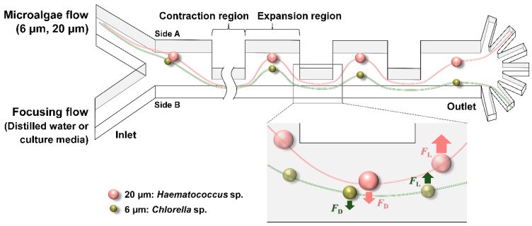
The microfluidic chip, essentially a tiny plumbing system, guides liquid through a spiral channel with repeating narrow and wide sections. Like leaves separating into different paths in a curving stream, cells experience forces that push them into different positions depending on their size. Computer simulations suggested that the largest particles would move toward the channel center while smaller particles remained closer to the inner wall.
Enriching Cancer Cells Without Chemical Labels
To test the chip, the researchers mixed MCF-7 breast cancer cells, a widely used lab-grown cell line, with white blood cells collected from healthy volunteers. Red blood cells were removed before testing, making this a more controlled sample than whole patient blood.
Across three experiments, the chip recovered 89.2% of cancer cells at its middle outlet and removed 86.9% of white blood cells. The cancer-cell share rose from about 9% in the material entering the device to 38% at that outlet. That is an important enrichment step, although the collected fraction still contained substantially more noncancer cells than cancer cells.
Conventional CTC workflows often rely on fluorescent dyes or antibodies to mark cells. Those labels can add preparation steps and may interfere with downstream work such as culturing cells or examining their genetic material. This system instead uses physical cell sorting first, then image analysis.
Teaching AI to Recognize Unstained Cells
After enrichment, the team used YOLOv8, a deep-learning system built to find objects within images, to distinguish cancer cells from white blood cells. The model analyzed brightfield microscopy images, the standard unstained images produced by many optical microscopes.
Fluorescent staining was used only to create a reference set for training. MCF-7 cells were stained green and white blood cells blue, allowing researchers to identify each cell type in matched images. The AI was trained using the corresponding plain brightfield images, so it learned to make predictions without requiring dyes during detection.
The researchers assembled 200 matched image sets, using 160 for training and 40 for validation. On prepared mixtures, the system produced 94.6% precision, meaning most cells labeled as cancer were cancer cells, and 94.6% recall, meaning it found most of the cancer cells present. Its specificity, the proportion of normal cells correctly excluded, was 95.4%.
Why This Matters
A blood-based CTC test could potentially offer a repeatable window into tumor behavior without surgery. If cells can be collected without harsh labeling procedures, they may remain more useful for later analyses, including single-cell sequencing or tests of how they respond to drugs.
Those possible uses remain unproven here. The researchers tested one breast cancer cell line in artificial mixtures, not the highly variable blood samples of people with cancer. They also evaluated the chip and AI modules separately rather than as one seamless automated instrument.
The next meaningful test is whether the platform can handle patient-derived samples, multiple cancer cell types, intact blood-cell backgrounds, and the far lower tumor-cell concentrations expected in real clinical settings. If it can, a chip that enriches cells physically and software that identifies them without stains could become a useful foundation for less invasive cancer monitoring.
Topics: Microfluidics · Cancer
Start here: Microfluidics explained
Related articles
- AI Chip Helps Find Rare Cancer Cells in Blood in New Liquid Biopsy Study
- Lab-on-a-Chip Enables “Liquid Biopsy” of Cancer Cells
- Using the polymeric circulating tumor cell chip to capture circulating tumor cells in blood samples of patients with colorectal cancer
- Groundbreaking Mass General-designed cancer cell-sorting chip brings liquid biopsies closer to clinic