Source: Cancer Medicine, by Saiying Ma; Xiaojia Wang; Peter Ping Lin; Lei Lei. AI-generated summary by biochip.com, published . Not independently reviewed.
Key takeaways
- CTC-Chip anti-EpCAM microcolumns reported 80% to 95% sensitivity and 90% to 97% specificity for detecting circulating tumor cells.
- T-μFS combined tetrahedral DNA frameworks, herringbone channels, and aptamers to achieve 80% to 90% sensitivity and 85% to 92% specificity for leukocyte-free isolation.
- No platform has emerged as a definitive clinical solution, and designs retain tradeoffs in capture efficiency, purity, visibility, cell release, and downstream analysis.
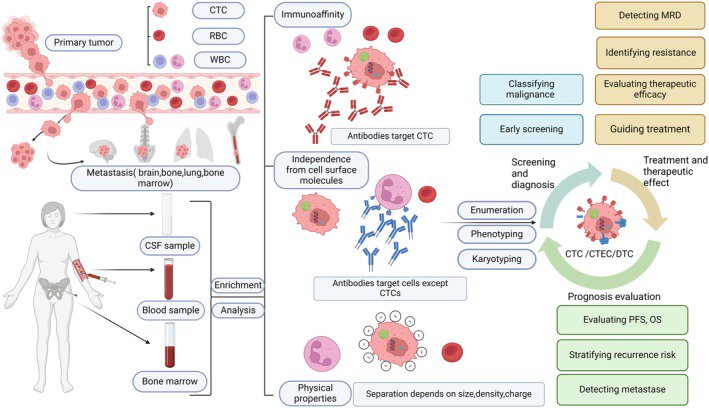
Circulating tumor cells, or CTCs, are cancer cells that break away from a tumor and travel through the bloodstream, making them a tantalizing target for a simple blood test in breast cancer. The article describes how newer microfluidic devices - tiny lab systems that steer fluids through narrow channels - are improving the odds of finding these rare cells among millions of normal blood cells. That matters because CTCs could help doctors do two hard things at once: track how a treatment is working and estimate how aggressive a patient’s disease may be. Older approaches often struggled with sensitivity, meaning they missed cells that were really there, or with specificity, meaning they risked mistaking other cells for tumor cells. The newer chip-based systems aim to solve both problems by controlling how blood flows, how tumor cells stick to a surface, and how captured cells are released for further study. In the source material, several platforms stand out, including the CTC-Chip, herringbone and graphene oxide chips, and a newer design called T-μFS that uses DNA nanostructures and aptamers, which are short molecules that bind specific targets like molecular Velcro. Across these examples, reported performance ranges are high enough to suggest real clinical promise, though each design still carries tradeoffs in visibility, capture strategy, and downstream analysis. Taken together, the story is less about one single gadget and more about a steady engineering push to turn a difficult biological signal into a practical tool for prognosis and treatment monitoring in breast cancer.
Why CTCs are so hard to find
Looking for CTCs is a bit like trying to spot a handful of specific grains of sand in a crowded beach. In a standard blood sample, tumor cells are exceedingly rare, while red blood cells and white blood cells dominate the scene.
That rarity is the central technical problem. A useful test must pull out the right cells without damaging them and without bringing along too many leukocytes, or white blood cells, that can muddy the readout.
How microfluidic chips improve capture
Microfluidic systems help because they do not just filter blood; they choreograph it. By controlling flow through tiny channels, engineers can increase the chances that a tumor cell will brush against a capture surface and stick, while unwanted cells keep moving.
The source highlights the CTC-Chip, which uses anti-EpCAM microcolumns. EpCAM is a protein often found on the surface of epithelial tumor cells, so antibodies against it act like selective hooks. In the cited reports, this platform achieved about 80% to 95% sensitivity and 90% to 97% specificity, strong numbers for a test trying to detect such rare targets.
The limits of early chip designs
But the first successful devices were not perfect. Microcolumn arrays helped with capture, yet their opacity made later analysis harder, especially when researchers wanted to closely examine the cells they had caught.
That matters because capturing a CTC is only step one. Doctors and researchers also want to characterize it - checking markers, comparing it with the original tumor, and asking whether it carries signs of drug resistance or metastatic potential.
Newer surfaces: herringbone and graphene oxide chips
To address those limitations, later devices shifted toward surface-capture designs such as herringbone and graphene oxide chips. A herringbone chip uses tiny groove patterns, like ridges in a washboard, to stir the blood as it flows past and improve contact between cells and the capture surface.
Graphene oxide chips take a different route by using a highly engineered surface that can support efficient binding and analysis. The broader idea behind both designs is simple: if you cannot increase the number of CTCs in blood, you can at least increase the chances that the rare ones will meet the right surface under the right conditions.
A DNA-based upgrade for cleaner isolation
One of the most interesting systems in the source is T-μFS, a microfluidic platform that combines tetrahedral DNA frameworks, herringbone channels, and aptamer-based capture. Tetrahedral DNA frameworks are nanoscale scaffold structures made from DNA, and they help present binding molecules in an ordered, efficient way.
The aptamers in this system serve as precise molecular binders, similar to antibodies but built from short nucleic acid sequences. According to the source, T-μFS achieved 80% to 90% sensitivity and 85% to 92% specificity for leukocyte-free isolation, an important step because cleaner samples make later analysis more reliable.
Why release and phenotyping matter
A good CTC platform should not trap cells forever. The source notes that postcapture enzymatic release can free cells after they are isolated, allowing researchers to examine them using markers such as CK and CD45.
These markers help distinguish likely tumor cells from blood cells. CK, or cytokeratin, is commonly used as an epithelial tumor marker, while CD45 is a standard marker for leukocytes; together, they provide a practical first pass at phenotyping, or classifying, the captured cells by identity.
What these systems could mean for breast cancer care
In breast cancer, a blood-based readout has obvious appeal. Tumors change over time, especially under treatment pressure, and tissue biopsies are invasive, limited to one site, and difficult to repeat frequently.
CTC detection offers a possible window into that evolving disease. If a patient’s CTC count rises, falls, or changes in character during therapy, clinicians may gain earlier clues about whether treatment is helping, failing, or selecting for more resistant cells.
Why This Matters
The source frames CTC detection as both a therapeutic and prognostic tool. Therapeutically, it could help monitor response in real time; prognostically, it may indicate how likely a cancer is to progress or spread.
What makes the microfluidic advances important is not just better engineering for its own sake. Better sensitivity and specificity mean fewer missed signals and fewer false alarms, which is exactly what a clinical test needs before it can guide decisions with confidence.
The road ahead
No single chip in the source emerges as the final answer, and that is probably the right takeaway. Different platforms solve different problems - capture efficiency, purity, visibility, release, or downstream analysis - and the future may depend on combining those strengths into workflows that fit real clinics.
Still, the direction is clear. As microfluidic design becomes more precise and cell analysis grows more sophisticated, CTC testing in breast cancer looks increasingly like a practical bridge between tumor biology and routine patient care, offering a less invasive way to follow the disease as it changes.
Topics: Microfluidics · Cancer
Start here: Microfluidics explained
Related articles
- Current Detection Technologies of Circulating Tumor Cells
- Circulating Tumor Cell Detection Technologies and Clinical Utility: Challenges and Opportunities
- Using the polymeric circulating tumor cell chip to capture circulating tumor cells in blood samples of patients with colorectal cancer
- The Liquid Biopsy Consortium: Challenges and opportunities for early cancer detection and monitoring