Source: Biomimetics, by Behnaz Eftekhari; Shawn P Grogan; Darryl D D’Lima (September 17, 2026). AI-generated summary by biochip.com, published . Not independently reviewed.
Key takeaways
- Scripps Health researchers reviewed multiscale vascular engineering, from centimeter-scale arteries to 10 to 15 micrometer capillaries.
- The review identifies microvascular organ-on-a-chip systems as high-fidelity platforms for studying vascular physiology, disease, and drug responses.
- The review does not establish clinical performance or long-term human outcomes for any vascular fabrication approach.
A review by Behnaz Eftekhari, Shawn P. Grogan, and Darryl D. D’Lima at Scripps Health lays out how engineers can build vascular networks that span from large arteries to tiny capillaries. The central challenge is not simply making hollow channels, but creating vessels that carry fluid, support living cells, respond to flow, and connect with surrounding tissue. The review evaluates microvascular organ-on-a-chip systems alongside scaffolds, cell-sheet constructs, decellularized matrices, and 3D bioprinting. Its practical message is that no single fabrication method can reproduce every part of the body’s vascular hierarchy.
Why Blood Vessels Are Hard to Rebuild
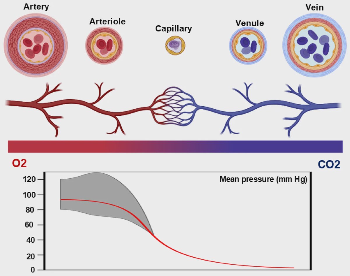
Natural blood vessels operate at dramatically different scales. The aorta measures several centimeters across, while capillaries, the smallest exchange vessels, are only about 10 to 15 micrometers wide. Each scale has a different job: large arteries withstand pulsatile pressure from the heart, while capillaries exchange oxygen, nutrients, and waste with nearby cells.
That difference creates a design problem much like building a transportation system that must include high-speed highways, local streets, and individual driveways. Engineered tissues need both larger conduits to deliver flow and dense microvascular networks to reach individual cells. Cells generally cannot survive more than 150 to 200 micrometers from a perfused vessel, making organized small vessels essential in thicker engineered tissues.
Cells and Flow Must Work Together
Endothelial cells, which line the inside of every blood vessel, are only one part of a functional network. Capillaries also rely on pericytes, support cells that help maintain vessel stability and barrier function. Larger vessels need vascular smooth muscle cells and fibroblasts to provide elasticity, contractility, and mechanical strength.
Flow is equally important. Blood moving across endothelial cells creates shear stress, a frictional force that influences cell alignment and helps maintain anti-clotting activity. The review notes that venous shear stresses typically range from 1 to 6 dyn/cm², while arterial values range from 10 to 20 dyn/cm². Channels that do not reproduce suitable flow conditions can develop endothelial dysfunction and a prothrombotic state.
Biofabrication Options Serve Different Scales
The review compares several approaches for constructing vascular systems. Electrospinning produces nanofibrous materials that resemble the extracellular matrix, the supportive material around cells. Decellularized matrices retain biological and mechanical cues from native tissues after their cells have been removed, while cell-sheet engineering builds dense, matrix-rich living layers.
3D bioprinting and sacrificial-template fabrication offer more direct control over channel geometry. In sacrificial-template approaches, engineers create a temporary structure within a material, then remove it to leave a hollow, perfusable channel. These methods can help shape vascular pathways, but the resulting networks still need appropriate cells, stable walls, and the ability to tolerate flow.
Microvascular Chips as Test Beds
Microvascular organ-on-a-chip models are a key part of the review's framework because they can recreate small, perfused vascular environments under controlled conditions. These systems use microscale channels to study human vascular physiology, disease processes, and drug responses. They can also help researchers test how endothelial cells and supporting cells behave under defined mechanical and biochemical conditions.
Such chips are not substitutes for complete vascular grafts. Instead, they function as high-fidelity experimental platforms for examining the rules that future engineered networks must follow. They can help researchers assess barrier behavior, flow responses, and cell interactions before moving to more complex tissue constructs.
Why This Matters
A usable vascular graft or engineered tissue must combine biology with physical design. Large-vessel constructs need sufficient burst pressure and mechanical compliance, meaning they can expand and recoil in a way that matches host vessels. Microvascular systems need fine patterning, controlled permeability, and reliable interactions between endothelial cells and pericytes.
The review argues for matching fabrication strategies to vessel-specific requirements rather than seeking one universal solution. Combining stem cell-derived vascular lineages, supportive cocultures, biomechanical conditioning, and multiscale fabrication could help create more stable and tissue-specific networks. The next step is to translate these design principles into perfusable systems that remain functional and integrate with host circulation.
Topics: Organ-on-a-chip · Heart & vascular health
Start here: Organ-on-a-chip explained
Related articles
- Engineering human multi-organ tissue chip niches for drug absorption, distribution, metabolism, excretion and toxicity prediction
- Setting out a roadmap for standardisation of organ-on-chip technology
- Mechanical Forces: The Hidden Conductor Shaping Organoids
- 10 Things to Know About Organ Chips and How They Benefit Humanity