Source: Biosensors, by Linyan Xie; Mengfei Wang; Xijia Luo; Shuoxian Xia; Qiongqiong Ren; Xuezhi Zhou (September 7, 2026). AI-generated summary by biochip.com, published . Not independently reviewed.
Key takeaways
- Henan Medical University researchers combined microfluidic light-scattering imaging with ResNet50 deep learning to classify individual lymphoma cells without fluorescent labels.
- The platform analyzed 800 single-cell patterns and achieved 94.75% average accuracy with a 0.986 average ROC area.
- The study tested only two fixed lymphoma cell lines, not patient samples or broader clinical lymphoma diversity.
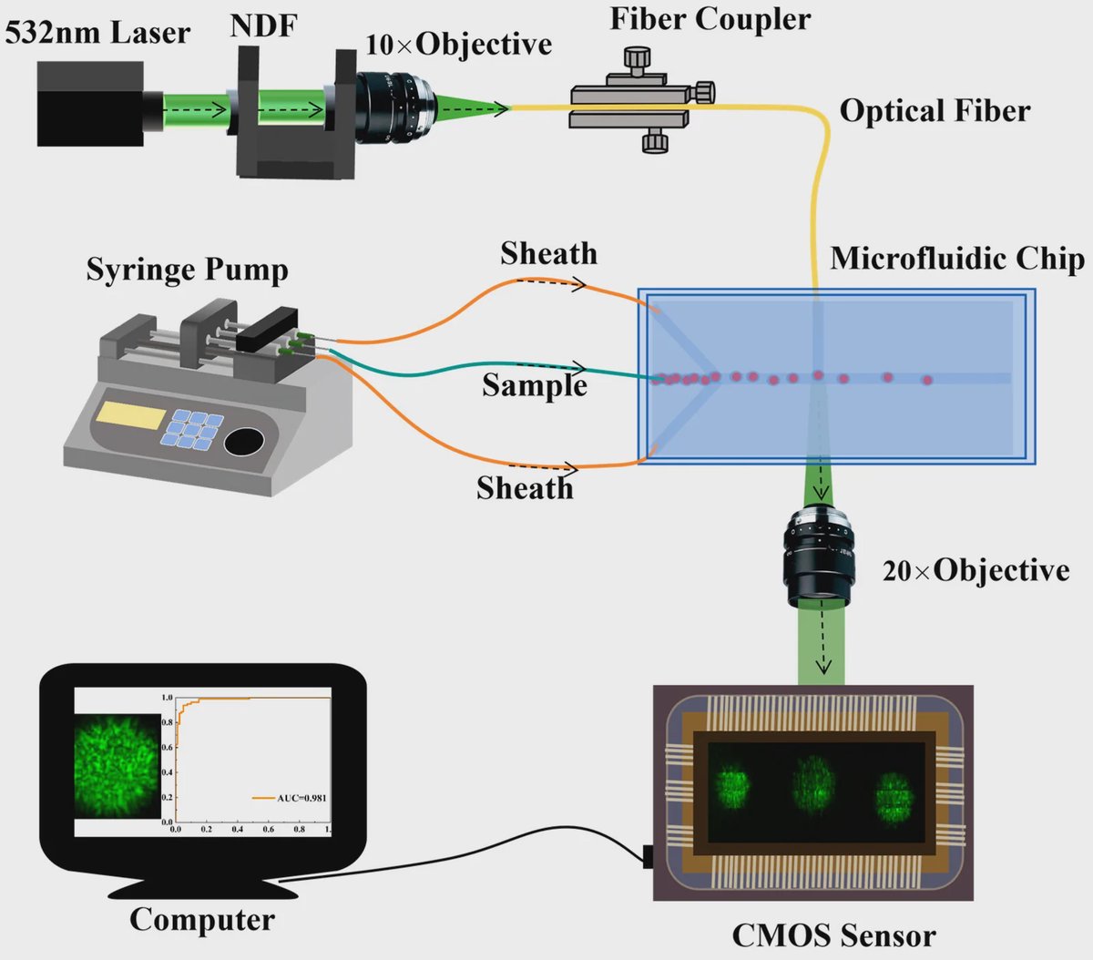
Researchers at Henan Medical University developed a microfluidic light-scattering imaging platform that distinguished two lymphoma cell lines without fluorescent labels. The system guides individual cells through a small chip, records their light-scattering patterns, and uses a deep-learning model to classify them. In tests involving 800 single-cell images, it reached an average accuracy of 94.75% and an average area under the receiver operating characteristic curve of 0.986. The work points to a way of extracting useful cellular information from light alone while reducing some of the preparation required by conventional cell-labeling methods.
Reading Cells Through Scattered Light
Cells scatter light differently depending on their shape, internal structures, and refractive index, which describes how light bends as it travels through a material. It is similar to shining a flashlight through differently textured pieces of glass: each redirects the beam in its own pattern. Here, those optical patterns served as label-free signatures for individual lymphoma cells.
The team tested human B lymphoma Daudi cells and human T lymphoblastic lymphoma SUP-T1 cells. Before biological experiments, it also used 4 micrometer silica microspheres and 6 micrometer polystyrene microspheres to confirm that the imaging system could reliably capture scattering patterns in flowing samples.
A Chip That Aligns Cells in Flow
The platform uses hydrodynamic focusing inside a microfluidic chip. This technique uses surrounding liquid streams to position cells in a narrow, controlled path, much like water currents steering a leaf into the center of a channel. Stable alignment helps the optical system measure one cell at a time as cells move continuously through the device.
The researchers collected two-dimensional light-scattering images, then automatically preprocessed the images before analysis. They designed the system to acquire scattering signals within a specific range, aiming to retain the cells' relevant optical information while reducing interference.
Deep Learning Finds the Patterns
Rather than asking researchers to manually select visual features from each scattering image, the team used transfer learning based on ResNet50, a neural-network architecture previously trained on large image datasets. Transfer learning adapts patterns learned in one image-recognition task to a new task, allowing a model to recognize subtle features with less task-specific data than training from scratch.
The dataset contained 800 single-cell scattering patterns. Using stratified five-fold cross-validation, which repeatedly trains and tests the model on different balanced subsets of the dataset, the system produced 94.75% average classification accuracy. Its average receiver operating characteristic area under the curve, a measure of how well a classifier separates two groups across decision thresholds, was 0.986.
Why This Matters
Lymphoma includes biologically diverse cancers of lymphocytes, so distinguishing cell types can inform diagnosis, disease classification, treatment decisions, and prognosis. Existing methods such as histopathology, immunohistochemistry, and flow cytometry can be highly accurate, but often involve staining, labor-intensive preparation, specialized instruments, and trained personnel.
By combining a microfluidic chip with optical sensing and automated image analysis, the platform offers a potential route toward continuous, single-cell measurements without added fluorescent contrast agents. The current results establish performance for two fixed lymphoma cell lines, not diagnostic performance in patient samples. Further studies will need to determine how the approach performs across the broader diversity of lymphoma cells encountered in clinical settings.
Topics: Microfluidics
Start here: Microfluidics explained
Related articles
- Microfluidic Impedance Biosensor Chips Using Sensing Layers Based on DNA-Based Self-Assembled Monolayers for Label-Free Detection of Proteins
- Machine Learning and Centrifugal Microfluidics for Bedside Sepsis Prediction
- Automated Microfluidic System for Point-of-Care Detection of Pathogens
- Microfluidic Co-Culture Platform for Taste Cell-Neuron Signaling