Source: MIT News, by Zach Winn (September 24, 2026). AI-generated summary by biochip.com, published . Not independently reviewed.
Key takeaways
- MIT and Johns Hopkins researchers built a handheld microfluidic device that collects viable cells from selected regions of excised tissue.
- The team grew organoids from cells collected from fresh human fallopian tube samples using controlled fluid shear.
- The device has been tested on excised tissue, not yet as an in-body diagnostic method for patients.
MIT researchers and collaborators at Johns Hopkins University have built a handheld microfluidic device that collects living cells from precisely chosen spots on excised tissue. In tests with human fallopian tube samples, the system recovered viable cells that could be grown into organoids, miniature lab-grown models of tissue. The work could give researchers a less destructive way to investigate the earliest changes linked to high-grade serous ovarian cancer, while preserving the original tissue for standard pathology.
A targeted way to collect cells
High-grade serous ovarian cancer, the most common form of ovarian cancer, often begins in the fallopian tubes. Its earliest precursor lesions can be microscopic, however, making them difficult to find and sample. That timing matters: the five-year survival rate can exceed 90 percent when ovarian cancer is caught early, but falls below half for stage 3 or 4 disease.
Conventional pathology preserves removed tissue in a chemical fixative, slices it into thin sections, and examines those sections under a microscope. This process is valuable for identifying suspicious tissue, but it kills the cells, preventing scientists from growing them or testing how they behave.
Kripa Varanasi's group at MIT wanted to collect cells from a specific location without disrupting the surrounding sample. Their device is designed to work on tissue after it has been surgically removed, an initial use case that could fit alongside existing pathology workflows.
Like a tiny, controlled rinse
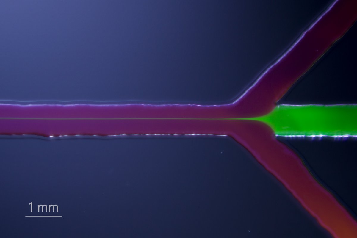
The device is a 3D-printed microfluidic tool, meaning it directs very small volumes of liquid through narrow channels. Think of it as a highly localized rinse rather than a brush or scalpel. One syringe creates a vacuum seal between the device and tissue, while a second syringe drives liquid across the selected surface.
That flowing liquid creates shear stress, the gentle dragging force produced when fluid moves across a surface. By controlling that force, the system detaches cells from a tiny targeted region without chemicals or enzymes. The collected cells then flow out for analysis or cultivation.
The team showed that it could adjust the applied shear stress for different cell types. Prostate cancer cells described as loosely attached could detach at 1 pascal of stress, while only a few bone cancer cells detached even at 5 pascals.
Keeping cells alive for further testing
The researchers compared their method with other cell-collection workflows and found that cells obtained with the fluidic approach remained viable and grew in culture more readily than cells detached by conventional methods. Viability is crucial because living cells can be expanded and studied in ways that fixed pathology samples cannot.
Using fresh human fallopian tube tissue provided by Johns Hopkins collaborators, the team collected living cells and grew them into organoids. These three-dimensional cell cultures can help scientists model disease and potentially test how cells from an individual patient respond to different treatments.
Rebecca Stone, a gynecologic oncologist at Johns Hopkins, collaborated with MIT engineers after being introduced to Varanasi by MIT professor Angela Belcher. Break Through Cancer supported the project, which is part of a broader effort to improve early ovarian cancer detection.
From excised tissue toward diagnostics
The initial plan is to use the device on tissue already removed from the body. Varanasi and colleagues say this could offer a more straightforward regulatory path and allow the technology to be integrated with routine histopathology, the examination of tissue for disease.
The group also envisions eventually adapting the approach to collect samples inside patients. Belcher's team is developing optical methods to identify suspicious tissue regions, and the cell-collection device could potentially retrieve viable cells from those exact locations for deeper study.
Why This Matters
Cancer testing often forces a tradeoff between preserving tissue for diagnosis and obtaining living cells for experiments. This microfluidic device aims to do both: sample a selected area while leaving surrounding tissue intact for pathologists. If further developed and validated, it could help researchers study how cancers begin in real human tissue and build more patient-specific disease models.
The device has so far been demonstrated on newly excised tissue, not as an in-body diagnostic test. The next steps will determine how reliably it can identify and sample suspicious regions, and whether those living-cell samples can contribute to earlier detection or personalized treatment decisions.
Topics: Microfluidics · Cancer · Diagnostics & precision medicine
Start here: Microfluidics explained
Related articles
- Microfluidic biosensors for biomarker detection in body fluids: a key approach for early cancer diagnosis
- The Liquid Biopsy Consortium: Challenges and opportunities for early cancer detection and monitoring
- Microchip Detection of Magnetically Labeled Cancer Biomarkers
- Microfluidic Point-of-Care (POC) Devices in Early Diagnosis: A Review of Opportunities and Challenges