Source: MethodsX, by Andrea Balsa-Díaz; Laura Vázquez-Vázquez; Elías Ferreiro-Vila; Lois Rivas-Meizoso; Antón L Martínez; José Manuel Brea; Mabel Loza; Santiago Pérez Rodriguez; Esteban Sinde; Ezequiel Alvarez; Bruno K Rodiño-Janeiro (September 22, 2026). AI-generated summary by biochip.com, published . Not independently reviewed.
Key takeaways
- Researchers standardized extracellular matrix coatings and flow conditions for Caki-1 kidney and A549 lung cells in organ-on-a-chip channels.
- The protocol uses PDMS microfluidic chips, coating materials, and peristaltic pumping to support adherent cells under controlled flow.
- The protocol does not establish performance across primary cells, other chip geometries, or clinical drug-testing applications.
A team from BFlow SL, the Universidade de Santiago de Compostela, and Hifas Innovation Hub has outlined a protocol for standardizing organ-on-a-chip culture under controlled fluid flow. The method focuses on Caki-1 kidney cells and A549 lung cells grown in microfluidic channels, where surface coatings and flow conditions can strongly affect whether cells attach and mature. It matters because small differences in chip preparation can alter cell behavior, making experiments difficult to reproduce across runs or laboratories. The protocol aims to make these flow-based cell models more consistent for biomedical research and drug development.
Why Standardization Is Needed
Most routine cell experiments still grow cells as flat layers in static dishes or multiwell plates. Those two-dimensional cultures are useful, but they do not reproduce the moving fluid, physical forces, and three-dimensional organization that cells experience in the body.
Organ-on-a-chip systems place cells inside small channels that can carry culture medium continuously. Like a tiny irrigation system for cells, the setup supplies nutrients while exposing cells to flow. That movement creates wall shear stress, the frictional force of fluid sliding over a cell layer, which can influence cell shape, metabolism, differentiation, and other biological responses.
Matching the Surface to the Cells
The team used chips made from polydimethylsiloxane, or PDMS, a transparent silicone material widely used in microfluidics. PDMS is biocompatible and gas permeable, but its water-repelling surface makes it difficult for cells to adhere without preparation.
To address that problem, the protocol evaluates extracellular matrix, or ECM, coatings for the channels. The ECM is the support material surrounding cells in tissues, and coating a chip with it gives cells biochemical and physical cues for attachment. The tested coating materials include collagen, gelatin, fibronectin, and poly-L-lysine, with several concentrations and solution conditions described for chip functionalization.
Two Cell Models Under Flow
The workflow uses Caki-1 cells, derived from kidney tissue, and A549 cells, derived from lung tissue. These cell lines provide two use cases for showing why a single chip-preparation recipe may not fit every cell type. The protocol emphasizes that ECM selection should be tailored to the cells being cultured.
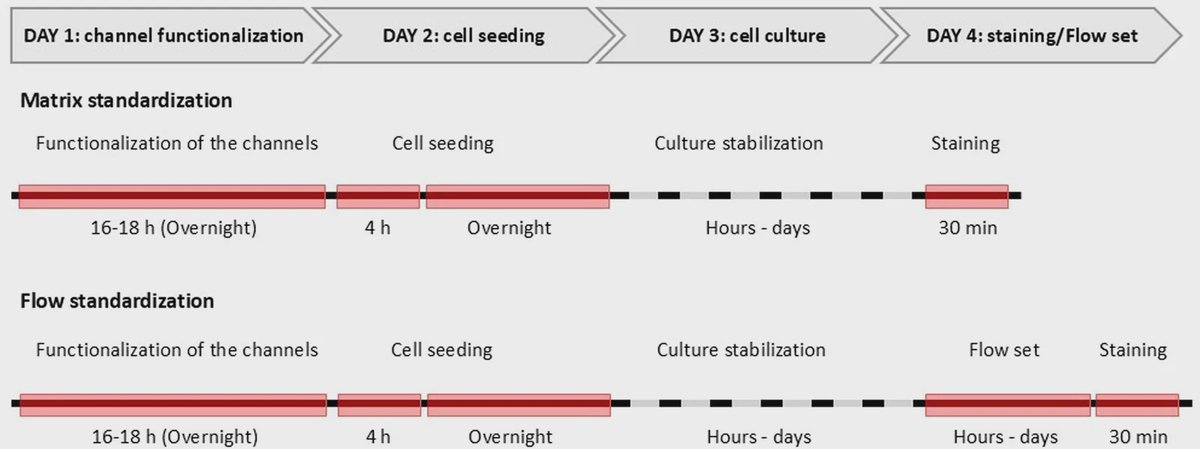
After coating the channels and seeding cells, researchers establish the biological microenvironment before applying controlled flow. That includes allowing sufficient time for cell adhesion and maturation, then using a peristaltic pump and tubing system to circulate medium through the chip. The experimental setup uses multi-linear channel chips, although the researchers state that the same overall approach can be adapted to other organ-on-a-chip designs.
A Practical Framework for Reproducibility
The protocol provides reagent preparations, equipment specifications, and steps for creating coating solutions. These details matter because seemingly minor changes, such as whether collagen is diluted in water or phosphate-buffered saline, can affect the interface where cells meet the chip.
It also separates two linked tasks: selecting a surface that can hold cells securely and defining flow conditions that model physiological forces without disrupting the culture. Together, those decisions determine whether a microfluidic model remains stable long enough to support an experiment.
Why This Matters
Organ-on-a-chip platforms are designed to bridge the gap between simple cell cultures and more complex animal studies. By combining cell culture with an engineered microenvironment, they can support three-dimensional structures, co-cultures, and dynamic perfusion in a controlled format. Better standardization could help laboratories generate more comparable data when using these systems to study disease biology or assess drug efficacy and toxicity.
The next step is to apply similarly explicit validation to additional cell types, chip geometries, and experimental questions. A reproducible starting protocol can make it easier to determine which differences in an organ-on-a-chip experiment come from biology, rather than from inconsistent chip preparation or fluid handling.
Topics: Microfluidics · Organ-on-a-chip
Start here: Microfluidics explained
Related articles
- Engineering Organoids and Organ-on-a-Chip Systems with Sensors
- Organoids, Organ-on-a-Chip, and the Case for Online LC–MS
- Organ-on-a-chip platforms for disease modeling and in vitro diagnostic applications
- TissueGnostics collaborates with Diya Biological Technology in organoid and organ-on-a-chip innovation